Waipapa Eyecare - Optometrists
  • Home
  • About
    • Meet the People
    • OCT - Optical Coherence Tomography
    • Zeiss Visufit 1000
    • 70% Off Replacement Lenses
    • Take a 3D Look around
  • Eye Health
    • Myopia - Nearsightedness
    • Hyperopia - Farsightedness
    • Astigmatism
    • Presbyopia
    • Dry Eye
    • Cataracts
    • Glaucoma
    • Macula Degeneration
    • Flashes and Floaters
    • Retinal Detachment
    • Macula Hole
    • Diabetic Retinopathy
    • Keratoconus
  • Eye Exams
    • Our Eye Exam Standard
    • Contact Lens Exams
    • Eye Exams For Kids
  • About Glasses
    • Frames
    • Frames & Face Shape
    • Lens Designs
    • Lens Options
    • Thinner Lenses
    • Cleaning Your Glasses
  • Contact
  • FAQ
    • Privacy Policy
  • Book Now
    • Become a Client

Macula Holes

Central vision eye condition

Macular Hole

A macular hole can affect central detailed vision, making reading, faces, and fine detail harder to see.

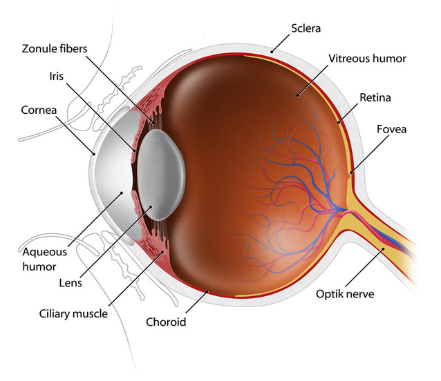
The macula is the central part of the retina at the back of the eye. It is responsible for sharp, detailed central vision.

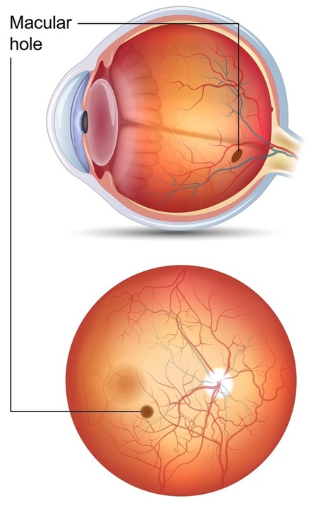
A macular hole is a small opening or gap that develops in the macula. It can cause central blur, distortion, a missing spot, or difficulty seeing fine detail.

Book Online OCT Eye Scans
Image showing a macular hole affecting the central retina A macular hole affects the central retina

New central distortion or missing vision should be checked promptly

Please phone us promptly if you notice new central distortion, straight lines looking wavy, a new dark or missing spot, sudden central blur, or a sudden change in reading vision.

These symptoms do not always mean a macular hole is present, but they should not be ignored.

What is the macula?

The macula is a small but very important part of the retina.

Detailed central vision

The macula helps you see fine detail, small print, faces, and objects you are looking directly at.

Reading and close work

Reading, sewing, computer work, and many detailed tasks rely on good macula function.

Colour and contrast

The macula also helps with colour, contrast, and sharp central vision.

A macular hole is different from macula degeneration

Both conditions affect the macula and central vision, but they are not the same. A macular hole is an opening or gap in the macula, while macula degeneration usually involves age-related retinal changes such as deposits, thinning, or leakage.

Macular hole image showing central retinal change

How a macular hole can affect vision

A macular hole affects the centre of the vision rather than the side vision. This can make detailed tasks difficult, even when peripheral vision remains useful.

Straight lines may look bent or wavy, words may appear blurred or missing, and a smudge, shadow, or blank area may appear near the centre of vision.

Symptoms may be more noticeable when checking one eye at a time, because the better eye can sometimes compensate when both eyes are open.

Possible symptoms of a macular hole

Symptoms can vary depending on the size and stage of the macular hole, and whether one or both eyes are affected.

Central blur

Reading, faces, television, or fine detail may become blurred or harder to see clearly.

Distortion

Straight lines, door frames, tiles, or text may look bent, wavy, or uneven.

Missing or dark spot

Some people notice a smudge, shadow, blank patch, or missing area near the centre of their vision.

Why macular holes develop

Macular holes are often related to age-related changes in the gel inside the eye, but there can be other causes.

Vitreous traction

The vitreous gel inside the eye can shrink and pull on the macula. Sometimes this pulling can contribute to a macular hole.

Age-related change

Macular holes are more common as people get older, although they can occur at different ages.

Other eye factors

Eye injury, high myopia, retinal conditions, or previous eye problems may also be relevant in some cases.

One eye can hide the problem

Because the other eye may compensate, some people do not notice a central vision change until they cover one eye at a time.

If one eye suddenly sees lines as bent, words as missing, or central detail as blurred, please arrange an eye examination promptly.

How a macular hole is checked

A full eye exam can help check whether symptoms are due to a macular hole, macula degeneration, swelling, vitreomacular traction, or another eye condition.

Vision and symptom history

We ask when symptoms started, whether one eye or both eyes are affected, and whether the change is sudden, gradual, stable, or worsening.

Macula examination

The optometrist examines the macula and retina for signs of structural change or other eye health concerns.

OCT imaging

OCT gives a detailed cross-section of the macula and is especially useful for checking the structure of the central retina.

Read about OCT eye scans

Our eye exam includes macula imaging

At Waipapa Eyecare, our complete eye exam includes retinal imaging and OCT. These help us check the macula and compare findings over time where needed.

If a macular hole or other significant macula change is suspected, we can explain what we have found and discuss referral to an eye specialist.

Read about our eye exam standard

If a macular hole is suspected

The next step depends on the OCT findings, symptoms, vision level, and whether the macular hole is early, partial, full thickness, stable, or progressing.

Monitoring

Some early or borderline findings may be monitored, especially if the vision is good and there is no full-thickness hole.

Referral

If a true macular hole is suspected, referral to an eye specialist may be recommended for assessment and advice.

Specialist treatment

Treatment, where appropriate, is usually managed by an eye specialist and may involve vitrectomy surgery.

Macular hole treatment is specialist care

If surgery is needed, the eye specialist will discuss the procedure, expected recovery, risks, benefits, and any positioning or follow-up instructions.

Surgery may help close the hole and improve or stabilise vision, but results vary depending on the size, duration, and other eye health factors.

Related eye health information

Central vision changes can have more than one cause. A complete eye exam helps check the bigger picture.

Macula degeneration

Macula degeneration can also affect central detailed vision, reading, and straight-line distortion.

Read about macula degeneration

OCT eye scans

OCT provides detailed 3D imaging that can help assess the macula, retina, and optic nerve.

Read about OCT

Flashes and floaters

Vitreous changes can cause floaters and flashes and may sometimes be linked with retinal traction.

Read about flashes and floaters

Is a macular hole the same as macula degeneration?

No. Both affect the macula and central vision, but they are different conditions with different management and treatment pathways.

Can a macular hole affect side vision?

A macular hole mainly affects central vision. Side vision is usually less affected, although the central blur can still be very disruptive.

Does every macular hole need surgery?

Not always. The best approach depends on the type, size, symptoms, vision level, OCT findings, and specialist advice.

Can glasses fix a macular hole?

Glasses may help if the prescription also needs updating, but they do not close a macular hole.

Concerned about central vision changes?

You can book online, phone us, or call in and see us at Waipapa Eyecare.

Monday to Friday: 9am - 5pm
Phone: 09 407 1065
Email: [email protected]
Book Online Contact Us

For urgent eye problems, sudden vision changes, flashes, floaters, eye pain, or injury, please phone us rather than using online booking.

Become a Client New Client

Waipapa Eyecare

Locally owned, independent optometry care in Waipapa. We provide thorough eye exams, modern diagnostic technology, quality lenses, and practical advice for your vision and eye health.

Book Online Privacy Statement

For urgent eye problems, sudden vision changes, flashes, floaters, eye pain, or injury, please phone us rather than using online booking.

Contact

Phone: 09 407 1065
Email: [email protected]
Hours: Monday to Friday, 9am - 5pm

Shop 8, 12 Klinac Lane
Waipapa, Kerikeri 0230

Quick links

  • Home
  • Our Eye Exam Standard
  • OCT Eye Scans
  • Zeiss Visufit 1000
  • Diamond Plus Offer
  • Privacy Statement
  • Contact Us
© 2021-2026 Waipapa Eyecare. All rights reserved.
Privacy Statement  |  Website content is general information only and does not replace personalised optometry advice.
  • Home
  • About
    • Meet the People
    • OCT - Optical Coherence Tomography
    • Zeiss Visufit 1000
    • 70% Off Replacement Lenses
    • Take a 3D Look around
  • Eye Health
    • Myopia - Nearsightedness
    • Hyperopia - Farsightedness
    • Astigmatism
    • Presbyopia
    • Dry Eye
    • Cataracts
    • Glaucoma
    • Macula Degeneration
    • Flashes and Floaters
    • Retinal Detachment
    • Macula Hole
    • Diabetic Retinopathy
    • Keratoconus
  • Eye Exams
    • Our Eye Exam Standard
    • Contact Lens Exams
    • Eye Exams For Kids
  • About Glasses
    • Frames
    • Frames & Face Shape
    • Lens Designs
    • Lens Options
    • Thinner Lenses
    • Cleaning Your Glasses
  • Contact
  • FAQ
    • Privacy Policy
  • Book Now
    • Become a Client